Fellowship-Trained Expertise
Retina & Diabetic Eye Care
Protect the light-sensitive lining of your eye. Dr. Adeel offers specialized diagnosis and clinical management of complex vitreoretinal conditions.
The Healthy Eye Clinic
Protect the light-sensitive lining of your eye. Dr. Adeel offers specialized diagnosis and clinical management of complex vitreoretinal conditions.
The retina is the thin, delicate tissue lining the inside back of the eye. It acts like the film in a camera, capturing light rays and translating them into neural signals that the brain recognizes as vision. The vitreous is the clear gel filling the space between the lens and retina.
Because retinal tissues cannot regenerate, diseases in this region pose a serious threat of permanent blindness. Dr. Adeel Ahmed Siddiqui has completed advanced medical and surgical vitreoretinal fellowship training, equipping him with the high-precision skills needed to manage retinal tears, diabetic complications, and macular degeneration.
We provide state-of-the-art care for systemic and age-related conditions affecting the posterior chamber of the eye.
Protecting Diabetic Sight
High blood sugar damages the microvasculature of the retina, leading to swelling (macular edema) or abnormal blood vessel growth (neovascularization) that can bleed into the vitreous. Frequent diabetic screenings are essential.
Preserving Central Vision
Age-related Macular Degeneration affects your central reading vision. We diagnose Dry AMD (cellular waste buildup) and treat Wet AMD (leaky vessels behind the retina) using highly effective intravitreal therapy.
Urgently Halting Vision Loss
A retinal tear occurs when the vitreous gel pulls away and tears the retina, which can lead to a full retinal detachment (a shadow or curtain over vision). This is a medical emergency requiring rapid intervention.
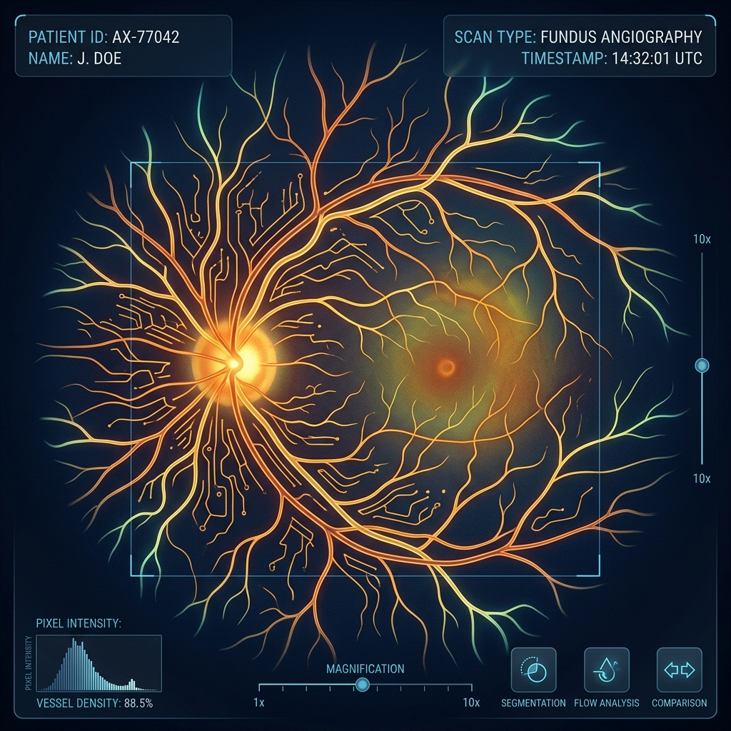
Retina care relies heavily on high-resolution imaging and targeted therapies. Depending on your diagnosis, Dr. Adeel may perform:
We administer dilating eye drops. It takes 20-30 minutes for your pupils to open fully, allowing Dr. Adeel to clearly inspect the entire periphery of your retina.
We take ultra-detailed scans of your macula and optic nerve. These scans measure retinal layer thicknesses at a micron scale, documenting any trace of fluid or swelling.
Dr. Adeel uses a specialized head-mounted light source and a handheld lens to manually examine the retina, looking for early vascular changes, tears, or thinning.
Dilating drops blur your reading vision and make you light-sensitive for 3 to 4 hours. You must bring sunglasses and have someone drive you home after your exam.